Diseases of the shoulder
Impingement
The term impingement comes from the English language and describes the trapping of anatomical structures under the shoulder roof. The impingement syndrome is a condition characterized by the association of specific symptoms: painful motion and stress-related pain, problems with overhead activities, pain when lying on the side, rarely pain at rest and constant pain.
The underlying causes can be vary, very often changes in the posture of and the position of the shoulder blade and an increase in the curvature of the thoracic spine lead to narrowing of the space under the roof of the shoulder (subacromial impingement). This can be aggravated by the formation of bone spurs and unfavorable congenital variants (acromion shape, lateral overhang of the shoulder roof over the humeral head, described by the CSA angle (critical shoulder angle) and the acromion index).
Advanced mechanical impingement can lead to the formation of a rotator cuff lesion .
The most frequently performed surgical therapy is endoscopic subacromial decompression (antero-lateral acromioplasty), in which the osseous constriction of the subacromial space is removed.


Rotator Cuff Rupture
Injuries to the rotator cuff (rupture) can result from direct or transmitted external forces. often falls on the outstretched arm or holding on to the railing when falling. The surrounding tendons of the humeral head (rotator cuff, consists of the subscapularis, supraspinatus, infraspinatus and teres minor) tear off from their attachment to the humerus. The result is the painful limitation of active mobility according to the affected tendons and reduced strength.
Today, in acute injuries tendons are usually refixated surgically (arthroscopically). Untreated ruptures have a tendency to become larger over time, which in turn leads to increasing impairment of biomechanical function. The final endstage disease is the development of a rotator cuff tear arthropathy (special form of secondary arthrosis), which often has to be treated with a reverse shoulder arthroplasty.
Rotator cuff lesions caused by natural aging processes are much more common than acute injuries . The individual course can be very different and is influenced by a variety of factors. This also includes the mechanical problems caused by the acromion shape and acromion spurs in impingement syndrome.


Frozen Shoulder
The adhesive capsulitis is a disease of the synovial membrane with various causes. The main symptom is a socalled frozen shoulder: the painful inability to move the arm also in small amounts.
The primary form (idiopathic, trigger unknown) is separated from the secondary forms for which an underlying cause can be described, e.g. accident, operation, radiation ...).
The disease has a phase-like course : in the 1st phase (freezing) the main symptom is the painfully restricted mobility. In the 2nd phase (frozen) the stiffness predominates, the pain is only secondary. In the 3rd phase (thawing) all adhesions will become soft and loose again.
The duration of the individual phases of the disease can vary greatly, and unfavorable courses can last for several years. Metabolic disorders (thyroid disorders) and insulin-dependent diabetes mellitus (IDDM) are particularly risk factors.
The therapy of choice in the early phase are corticosteroids followed by physiotherapy, exercise treatment and personal exercises. Unfavorable courses (refractory to therapy) sometimes have to be treated surgically.


Calcific Tendonitis
The calcific tendonitis is a condition in which calcific deposits can form in the tendons of the rotator cuff and bursa for unknown reasons. The disease has a phase-like course, the spontaneous regression and dissolution of the deposits is the rule, which can sometimes take several years. The supraspinatus tendon is most commonly affected.
There is a particular risk in the case of metabolic disorders (thyroid disorders) and the combination with an impingement syndrome.
The therapy of choice in the early phase is physiotherapy and personal exercises. The aim is the functional expansion of the subacromial space (sliding space of the humerus head under the shoulder roof).
The shock wave therapy (ESWT) can stimulate the release of calcium deposits in some cases.
Unfavorable courses (refractory to therapy) sometimes have to be treated surgically. The calcium deposit is removed mechanically with an arthroscopic operation. .


Shoulder Instability
The shoulder joint (gleno-humeral joint) is the most flexible joint in the human body. The very large range of motion makes special demands regarding stability, which is not provided by the bony anatomy alone, but is an interplay of static and dynamic factors.
A stable joint with a full range of functions is the result of the dynamic interaction of all local actors and the central nervous system with the movement control and thus comparable to a symphony orchestra. If one of the main instruments in the orchestra is damaged or one of the musicians does not play well enough, the overall picture (the harmonic sound of a symphony) will be compromized.
The most important static stabilizer is the capsule-labrum complex with the labrum and the joint capsule and its reinforcing ligaments (glenohumeral ligaments). The humeral head rests on the anterior and posterior lower band (aIGHL and pIGHL) like in a hammock.
Traumatic Dislocation
A dislocation of the shoulder joint can occur due to external force in the context of an accident, in the majority of cases the direction of the dislocation is forward (anterior). Young male adults are particularly at risk. Medical intervention in the emergency room is often necessary, the joint is "reduced" (repositioned), sometimes with a short anesthetic intervention.
As a result of the dislocation, the capsule-labrum complex is often so severely injured (Bankart lesion), that surgical refixation becomes necessary (arthroscopic Bankart repair) .


The enganging of the humeral head on the front edge of the glenoid during the dislocation can result in a bony impression ( Hill-Sachs lesion ) on the rear upper humeral head, which in some cases can be so large, that the head repeatedly tends to engage and dislocates. In these cases, surgical intervention is necessary. A so-called remplissage is often carried out, in which the capsule and tendon attachment of the infraspinatus are fixed into the defect in such a way, that it is no longer possible to mechanically engage it (door stopper effect).
Bony Bankart's Lesion
Traumatic shoulder dislocations can lead to a fracture of the anterior edge of the glenoid (bony Bankart lesion, Bankart fracture, bony Bankart). In the case of acute and large fragments, osteosynthesis is promising (screw fixation of the fracture, open or arthroscopic).
Small fragments can be successfully stabilized with arthroscopic suture anchor refixation in conjunction with the capsule-labrum complex.


Glenoid Bone Defect
Improperly healed Bankart lesions or recurrent dislocations can lead to bone defects in the anterior glenoid. The consequence is a mechanically caused recurrent instability. The combination of this clinical picture with a significant Hill-Sachs lesion (engaging Hill-Sachs lesion) is particularly unfavorable (so-called "off-track lesion").
The bone defect on the socket can be refilled with a bone chip (arthroscopic or open).
As a very reliable alternative, especially with regard to risk of recurrence of a dislocation, the coracoid transfer according to Latarjet has been proven to be very reliable.
Posterior Shoulder Instability
Dislocations of the humeral head to the rear occur more frequently in epileptic seizures and electrical accidents . It is not uncommon for posterior instabilities to occur in patients with hypermobility syndrome and multidirectional instabilities (MDI).
Posterior instabilities are often clinically overlooked or misdiagnosed.
The therapy usually is possible with arthroscopic stabilization, the techniques are comparable to those of the anterior instability (Bankart repair, bone block, capsule tightening).
Multidirectional Instability (MDI)
Patients with instability in several directions ( MDI ) often have a pronounced "connective tissue component", i.e. there may be disorders of the collagen metabolism, some of which can also be hereditary (Ehlers-Danlos syndrome) or simply very "soft joints" as a variant of normal anatomy. A clinical feature is often a pronounced laxity of the shoulder joints (also known as hyperlaxity ), which can be detected with various tests.
The therapy of choice initially is conservative treatment with improvement of the scapulo-thoracic rhythm and neurophysiological control of the muscles and improvement of the sequences of movements.
However, often surgical stabilization becomes necessary in the course of the disease, in which arthroscopic circumferent capsular repair (capsular shift) with a reduction of the joint volume is the treatment of choice.


AC-joint Dislocation
The AC-joint (acromio-clavicular joint) connects the collarbone (clavicula) with the roof of the shoulder (acromion). The joint is very firm and not very flexible, but not completely rigid. The joint capsule which is also known as the acromio-clavicular ligament and is very firm, especially at the back. Two very firm ligaments (trapezoid and conoid, cc ligaments) connect the clavicle with the coracoid process. The third pillar of stability for the AC-joint is the fascia of the trapezius, deltoid, and pectoralis major.

Injuries to the AC joint often occur as a result of direct or transmitted violent force to the shoulder, typical are falls on the shoulder with the arm at the side, falls while skiing or mountain biking ... Many patients even hear a loud noise, the shoulder is painful and sometimes immediately the relative elevation of the collarbone is palpable and visible.
Incomplete lesions (Rockwood I and II, partially III) are treated conservatively. Advanced instabilities (Rockwood IV, V, VI, VII) or those with additional horizontal instability components are treated surgically in a timely manner .
There is a large number of surgical procedures, modern arthroscopic-assisted pulley systems (e.g. tight-ropeTM ) allow anatomical healing and early functional follow-up treatment with very good long-term results.
If the correct time for the operation is missed or if chronic AC-joint instability has developed for other reasons , the use of additional tendon material becomes necessary; most often, the gracilis tendon from the lower leg is used, as in a cruciate ligament surgery.
Treatment of actue lesions

Chronic instability with
autologous gracilis tendon

Degenerative AC-joint
Age-dependent joint wear of the AC-joint occurs very often in the middle of the lifespan and becomes painful ( symptomatic AC-joint osteoarthritis ). In the early stages, conservative therapy is the treatment of choice, injection treatment is often applied.
Endoscopic AC-joint resection (lateral clavicular resection) can be performed if pain is refractory to therapy. With special rotating instruments (acromionizer), the bone of the collarbone removed under an endoscopic view in order to restore the joint space.


SLAP Lesion
Lesions of the upper biceps tendon anchoring complex ( superior labrum anterior posterior ) often occur with traction injuries to the arm, but can also result from chronic overload damage in overhead sports (baseball, volleyball, handball ...) .
Combination injuries associated with shoulder dislocations are quite common.
Spontaneous healing is practically impossible due to the unfavorable anatomical conditions.
SLAP lesions are often overlooked or underdiagnosed , even on MRI; the thorough taking of patient histor and clinical examination with special tests are decisive.
For therapy-resistant complaints, either SLAP repair (young patients, traumatic events, combined injuries with shoulder dislocation) or tenodesis of the long biceps tendon in the sulcus ( suprapectoral LHB tenodesis ) are possible.
SLAP lesion
SLAP repair
LHB tenodesis


Cartilage Lesions
The hyaline joint cartilage is a unique tissue that enables almost friction-free movement between the joint partners. Very few cartilage cells create the biomechanical prerequisites for these excellent properties through the production of special molecules and fibers (collagen, glycoproteins, elastane ...).
The regenerative capacity of the cartilage tissue is very limited, both for single impact loads and for repetitive overload.
Once the cartilage has been damaged, the ultimate goal is to prevent progression, to preserve the joint and to prevent the downward spiral of osteoarthritis. In addition to conservative procedures (e.g. hyaluronic acid injections ), surgical measures can also be considered (e.g. cartilage smoothing).
Cartilage repair procedures are reserved for specific defects and young patients and are the exception (e.g. autologous chondrocyte transplantation or implantation - ACT / ACI, osteochondral transplantation - OAT, etc.).
Nano-fracturing

ACI

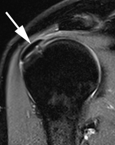
High-resolution MRI 6 months after OAT at the upper humeral head (right dGEMRIC-MRT)


Osteoarthritis
The age-related degenerative wear of the shoulder joint (primary omarthrosis) leads to typical changes of the anatomy: joint space reduction, osteophyte formation, deformation of the humeral head, decentering of the joint. Clinically, this is associated with painful restriction of movement, crepitation and, very often, pain at rest and at night. In the final stage, the therapy of choice is the replacement of the joint.
On the one hand, the right time for the operation is determined by the patient and his / her complaints. From a medical point of view, there are situations that are decisive in terms of therapy and the chances of satisfaction after a successful operation: the extent of the joint stiffness and shortening of the tendons and ligaments and the degree of the bony changes (advanced glenoid erosion and decentering can often only be treated with bone augmentation and and a reverse arthroplasty.
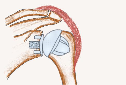
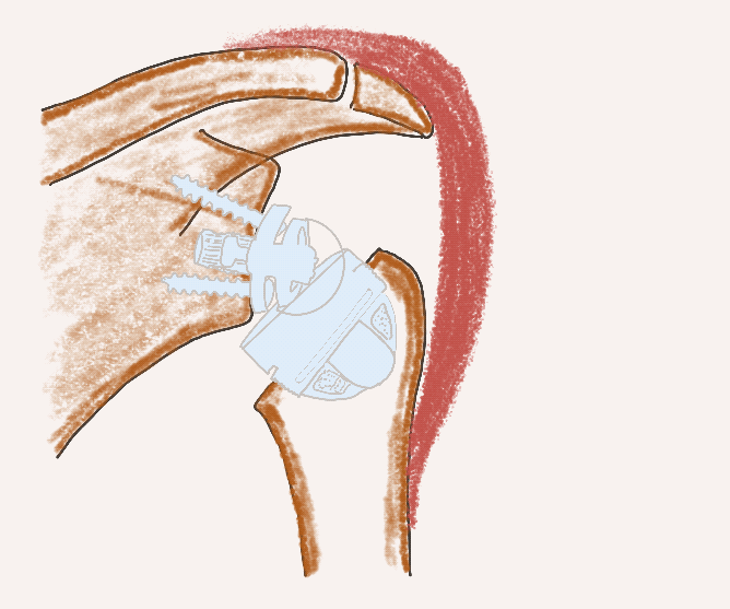
In general, the following rules apply regarding surgical therapy: as gentle on the soft tissue, bone-saving and anatomically precise as possible, with an anatomical implant (total shoulder arthroplasty) if the rotator cuff is preserved.
Modern prosthesis systems can be implanted without a classic stem and are individually adaptable and modular, and parts can be changed and, if necessary, converted into reverse systems.
Stemless anatomical implant system (TSA)
Stemless reverse implant system (RSA)
Humerus Fracture
Broken bones in the upper arm are one of the most common types of fractures. In young patients, the causes are serious accidents, in older patients there are often only falls at home. The reduced bone quality (aging and osteoporosis), especially in elderly women, present a significant challenge to the specialist.
Many proximal humeral fractures can be treated conservatively: temporary immobilization and early functional therapy.
The goal of surgical therapy is to allow the fracture to heal and to protect the soft tissues. Locking plate systems are used successfully to achieve this.
Primary reverse fracture endoprosthesis (reverse shoulder arthroplasty) has become the treatment of choice for comminuted fractures, cases of reduced bone quality and advanced age.


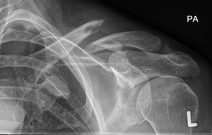
Clavicle Fracture
Fractures of the collarbone often occur after serious accidents with direct or transmitted external violent force. The cause can be, for example, a skiing accident, a fall from a mountain bike or motorcycle.
Many fractures are only slightly displaced and stable and therefor can be treated conservatively.
Displaced and comminuted fractures are problematic, especially those that are close to the AC-joint and thus have the potential to impair the stabilizing ligament complex. In these cases, the therapy of choice is osteosynthesis with a modern locking plate system.
The final healing often takes a comparatively long time, but has a good prognosis.