Surgical Procedures
Arthroscopy

Cartilage damage to the glenoid


Arthroscopy is a modern, minimally invasive therapy method in which operations in the joint are carried out using the so-called "keyhole technique" without the classic opening of the joint (arthrotomy).
This is made possible by the use of small optics (Hopkins rod lens system) and special arthroscopic instruments that are introduced through stab incisions. The anatomical and diseased structures can be visualized in zoom mode and addressed very specifically on high-resolution monitors.
The advantage: less damage to the structures surrounding the joint, better visualization, more precise work and better rehabilitation of the patient.
Over the years, special techniques and instruments have developed that have revolutionized treatment, such as the circumferential capsulorrhaphy for multidirectional instabilities (MDI), SLAP repair, endoscopic-assisted AC-joint stabilization and much more.
The joint is filled with saline solution and kept absolutely anemic with light pressure using pump systems. Even the smallest bleeding spots can be stopped immediately with special ablation probes, another advantage of the minimally invasive technique.
The risk of complications is very low and of course also depends on the operation performed.
Nowadays, even the most complex muscle flap procedures and other technically complex interventions can, in some cases, be successfully carried out arthroscopically. A trend that continues.

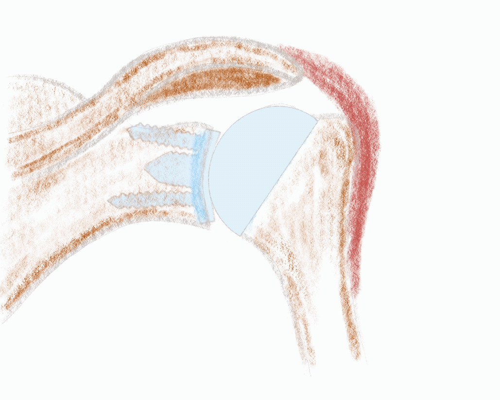
Intact SLAP region

Intact SSP tendon

Subacromial decompression
Open Procedures
Nowadays, surgical techniques that are not amenable for arthroscopicy or disadvantageous are still carried out openly. Classic open procedures are the Joint replacements and often complex reconstructions and revision operations .
When choosing the surgical procedure, it is particularly important to have the best possible prospect of achieving the surgical goals with the best possible result for the patient. The length of the skin incision is of secondary importance.
Even open procedures are now in many cases minimally invasive in the sense that as few soft tissues as possible are injured during the operation. This is achieved primarily through careful OR planning and the choice of suitable approaches.
Classic procedures, which are still mostly carried out openly today, are muscle-tendon transfres ( pectoralis major transfer PMT , Latissimus dorsi transfer LDT, coracoid transfer to Latarjet among others).
With the combination of small open approaches and endoscopic procedures, the advantages of both methods can often be sensibly combined, for example with endoscopically assisted AC joint stabilization .

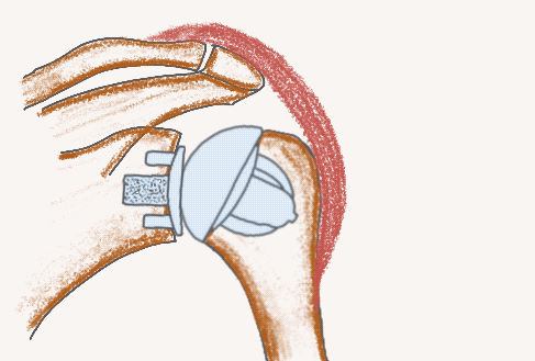
Endoscopically assisted ACG stabilization

Pectoralis major transfer (PMT)

Latissimus dorsi transfer (LDT)
Joint Replacement
Nowadays, joint replacement is almost exclusively carried out through a small anterior delto-pectoral approach. The soft tissues are treated very gently and structures that have to be detached (such as the tendon of the subscapularis muscle) are carefully and very stable refixated.
In the follow-up treatment, consideration must be given to the healing of the soft tissues with individual follow-up treatment plans for each patient.
Total Shoulder Arthroplasty TSA
A functioning shoulder joint needs intact soft tissue structures for powerful movement. The rotator cuff in particular must be intact and functioning so that the joint moves stable and centered. This is also the case after the implantation of an anatomical shoulder endoprosthesis. The worn off surfaces are replaced and the joint play is restored through perfect soft tissue balancing.
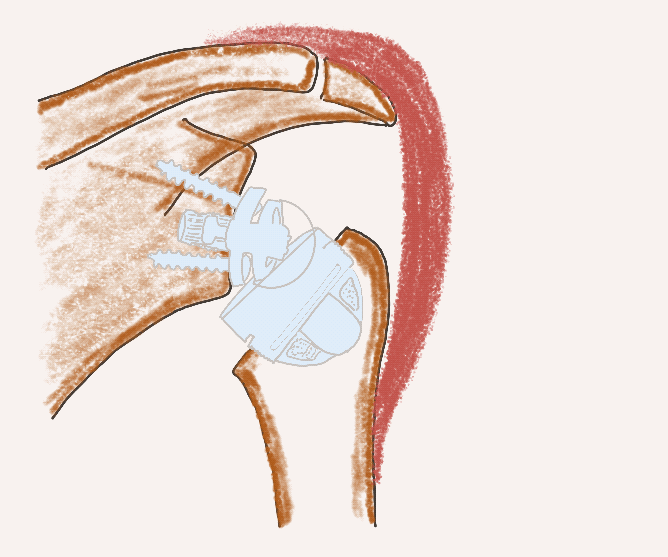
Reverse Shoulder Arthroplasty RSA
If the rotator cuff is severely damaged, the joint is very unstable or the muscles are no longer capable of performing (muscular atrophy and fatty degeneration), the reverse shoulder endoprosthesis is used. The spherical surface (glenosphere) is now on the scapular side (glenoid) and the cup on the humerus head. The two joint partners are now, in contrast to the natural joint, very conformous and highly stable. This mechanism and the changed geometry can compensate for the lack of function of the rotator cuff. The new powerful motor of the shoulder is now mainly the deltoid muscle, which must therefore be intact.

Stemless TSA and RSA
Both anatomical shoulder endoprostheses (TSA - total shoulder arthroplasty) and reverse shoulder endoprostheses (RSA - reverse shoulder arthroplasty) can now be inserted using modern stemless implants. The prerequisite is good bone quality at the upper arm.
Advantages of the stemless implants: bone-saving surgery and improved positioning of the implants regardless of the shaft axis. As a result there are improved options for revision surgrey and an optimal implant position.
The modern implant systems are convertible, so that only individual parts can be exchanged during a revision, if necessary.
stemless anatomical endoprosthesis
Stemless TSA
stemless reverse endoprosthesis
Stemless RSA
Tendon replacement
TEAR patch
Rotator cuff lesions are common in the population and increase with age. Surgical treatment of such tendon tears is one of the most frequently performed operations at the musculoskeletal system.
How can the progression of chronic, massive and irreparable rotator cuff lesions to cuff tear arthropathy be prevented? Is there a solution for treating irreparable rotator cuff tendon defects to restore function without pain?
For people of working age there are muscle-tendon transfers such as the pectoralis major transfer (PMT) for tears in the subscapularis and the latissimus dorsi transfer (LDT) for tears in the supra- and infraspinatus tendons. The LDT, in particular, as a complex operation, is still carried out successfully in specialized centers today.
In recent years, efforts have increasingly been made to bridge the tendon defect with tendon replacement materials (interposition) or to reinforce tendon sutures with such materials (augmentation). The use of commercially available specially prepared patches from the skin of donors is already possible arthroscopically with good short and medium-term results in special centers, especially in the USA.
In Germany, patches made from animal donor tissue or human skin from deceased donors have so far been available almost exclusively. With all of these materials, there remains a small residual risk of infection. But what is even more important, immunological reactions in the sense of rejection or intolerance must be expected in some patients, after all, the tissue is foreign to the body. The biomechanical properties of the known patches are usually not as good as those of normal tendon tissue, so that a real tendon replacement (interposition) is not permitted, but only a reinforcement of the suture (augmentation).
An ideal tendon replacement patch can be made from immunologically and infectiologically harmless endogenous material. A high elasticity and strength should allow immediate resilience after healing and the transfer of the forces that occur with powerful movement.
One promising option is the harvesting of the semitendinosus or gracilis tendon (so-called hamstring tendons) from the lower leg. In analogy to the treatment of patients with cruciate ligament rupture (approx. 75,000 operations per year in Germany), these tendons can be obtained in a minimally invasive manner with little donor site morbidity. The biomechanical tests showed a more than sufficient strength of the patch compared to the healthy rotator cuff.
The TEAR patch is made on a frame by alternating over and under weaving (comparable to a small patch of carpet). The tendons are knotted or sewn together. The finished patch is then sewn into the tendon defect of the rotator cuff during the operation and anchored to the humerus with suture anchors or transosseous sutures.
This treatment method was developed at the ATOS Clinic Fleetinsel Hamburg in cooperation with partners from the University Medical Center Hamburg-Eppendorf and is completely new. However, the theoretical foundations and principles have long been known and researched. Nevertheless, it must be emphasized that this is an attempt for treatmentment for which there is insufficient data about the success of the therapy. However, in the event of a complete failure of the therapy (tearing of the patch, lack of functional recovery of the shoulder, etc.), most of the alternative therapeutic methods (latissimus dorsi transfer, reverse shoulder arthroplasty ...) could still be carried out.


TEAR patch in holding device after biomechanical testing> 1,000 N
Albers S, Ono Y, Kirchner F, Fal MF, Kircher J. Midterm outcomes of autologous bridging of rotator cuff tears with an autologous tendon patch (TEAR patch). J Shoulder Elbow Surg 2024;33-2:312-20.